
Lipomyelocele is one of the most common closed spinal birth defects. It is seen in the thoracolumbar junction (the part of the spine that connects the thoracic spine to the lumbar spine) and usually presents as a fatty subcutaneous mass.
What is Lipoma?
A lipoma is a non-cancerous, fatty lump that grows under the skin. Lipomas are soft, round or oval-shaped, and usually move easily when touched.
What is Lipomyelocele?
Lipo = fat
Myelo = the nerves of the spinal cord
Cele = a swelling
A Lipomyelocele is a type of lipoma that occurs with Spina Bifida. A Lipomyelocele is a fibrous fatty tissue mass that develops in the spinal column and extends on the backside through a Spina Bifida defect (gap or opening in the malformed spine). A Lipomyelocele is located under the skin and it connects the spinal cord against the skin. The defect is covered by the skin which makes it “hidden” (occult) or closed Spina bifida (incomplete closure of the neural tube). Lipomyelocele is the most common type of occult SB and often occurs in the lowest part of the spine.

Image Description
Posterior lipomyelocele. (A) Image showing a skin-covered swelling in the lumbar region. (B) T2 axial and (C) T1 sagittal images and illustration (D) showing an intraspinal lipoma herniating into the subcutaneous fat through a posterior spina bifida. Case courtesy of Amarnath Chellathurai of Stanley Medical College
What is the difference between Lipomyelocele and Lipomyelomeningocele?
Lipo = fat
Myelo = the nerves of the spinal cord
Meningo = the meninges, or membranes (coverings) around the spinal cord
Cele = a swelling
While both “Lipomyelomeningocele” and “Lipomyelocele” refer to spinal cord malformations involving fatty tissue (lipoma), a key difference is that in a Lipomyelomeningocele, the fatty mass extends outside the spinal canal, whereas in a lipomyelocele, the fatty mass is located within the spinal canal, meaning a Lipomyelomeningocele is considered a more severe form of the condition; essentially, the “meningo” part indicates involvement of the meninges (the protective membranes around the spinal cord) which are not involved in a Lipomyelocele.
When does it occur?
It occurs during the third week of pregnancy until the end of the eighth week of pregnancy (embryonic stage of development), often before a woman even knows she is pregnant.
What are the causes?
A Lipomyelocele is caused by premature separation of the superficial ectoderm (which forms skin) from the neural ectoderm (nerve cells) which forms the spinal cord.
In simple words, Lipomyelocele is caused by a defect in the closure of the neural tube during early embryonic development. This defect allows mesenchymal cells (muh·sing·kuh·muhl selz) (MSCs) to enter the neural tube, which prevents it from closing properly. The mesenchymal cells then develop into fat cells, forming a fatty growth called Lipomyelocele. The Lipomyelocele attaches to the spinal cord and its membranes, causing tethering of the spinal cord. The tethering can be symptomatic causing Tethered Cord Syndrome.
What are the symptoms of Lipomyelocele?
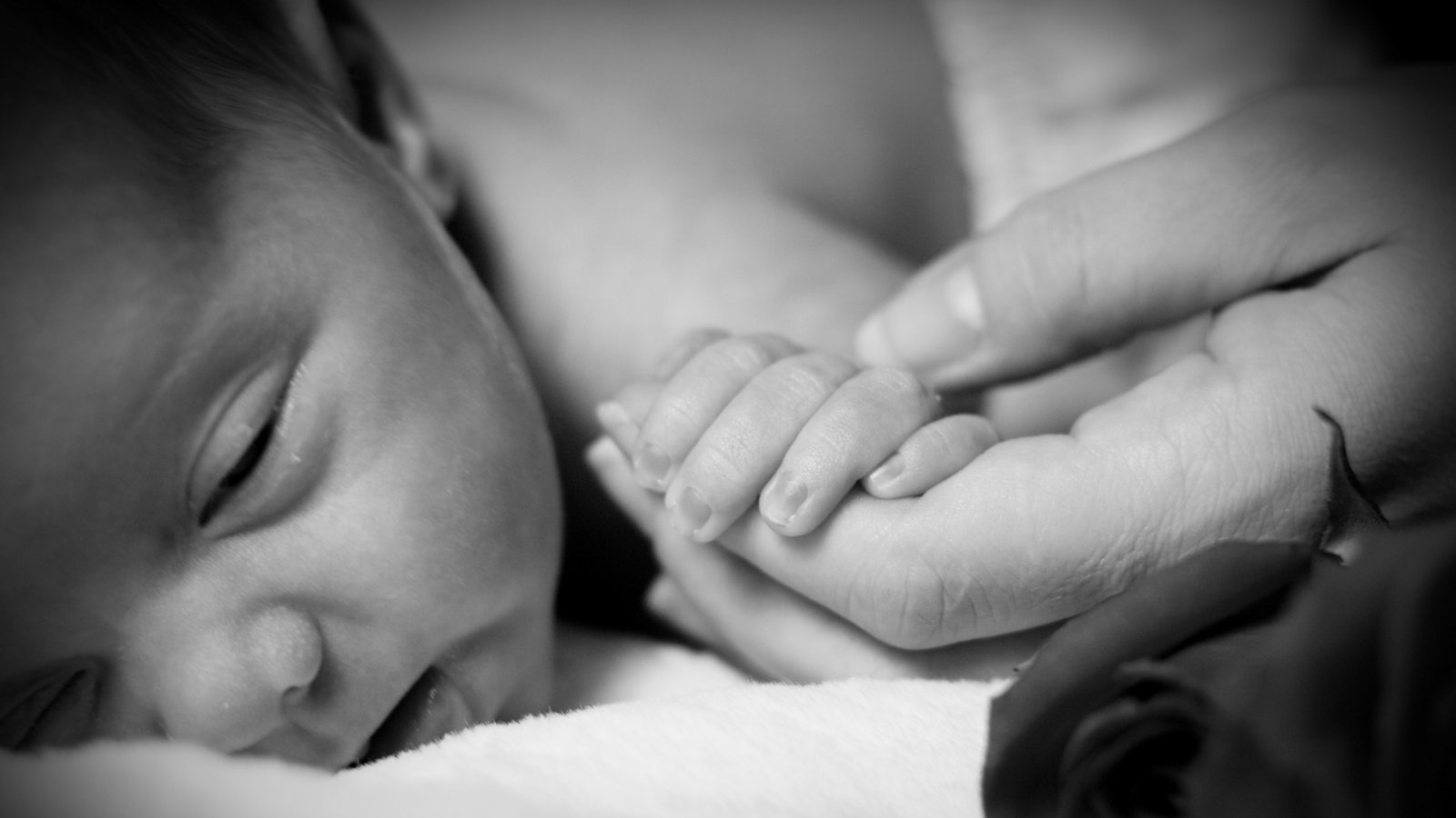
As the baby grows, urinary and bowel incontinence can occur. In addition, poor or absent sensation in the lower part of the body, limb weakness, difficulties with walking, and pain might be observed. As spoken earlier in our articles, the location of the Lipomyelocele will dictate the symptoms.
What is the treatment for Lipomyelocele?
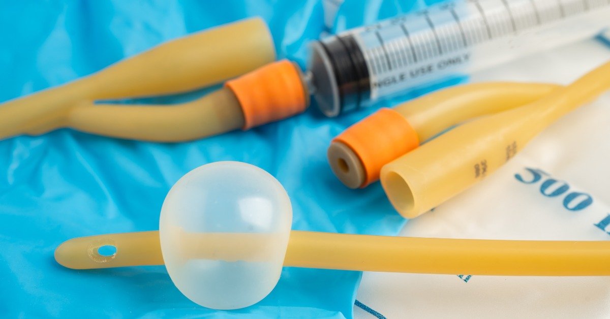
Treatment varies, initially diagnosed through clinical evaluation, followed by, a series of scans and tests to confirm the diagnosis. These tests may include an X-ray, a Magnetic Resonance Imaging (MRI) scan, coupled with additional tests like Urodynamic Studies. Please note that MRI is better than CT to evaluate Lipomyelomeningocele.
Tethered Cord Syndrome (TCS) is rarely an issue for children, especially with Myelomeningocele and is usually asymptomatic. However, in rare instances, these children may develop symptoms as they grow. The tension, or stretching, of the spinal cord, due to the increased length of the spine can lead to progressive neurological, urological, or orthopaedic deterioration. Sometimes, de-tethering surgery may be needed. In our opinion, de-tethering surgery should not be repeated as more scar tissue will develop causing re-tethering and other problems. Please read our article on de-tethering and Tethered Cord Syndrome.
Every Spina Bifida case is unique, and after individual assessment, the best treatment plan can be devised. You will have to consult specialists like a Neurosurgeon, an Orthopaedician, a Gastroenterologist and a Urologist.
Please Note
The information provided on our website is not intended as medical advice for any individual. Since specific cases may differ from the general information presented, SASHA recommends consulting a qualified medical or other professional for personalized advice.
About the Author

Raul/DJ Vivek
Meet Vivek Bharadwaj, a remarkable individual who has defied the odds and soared to new heights despite living with Spina Bifida. As the founder of the Sasha Foundation, Vivek tirelessly advocates for others facing similar challenges. His unwavering commitment to support, awareness, and empowerment had made a lasting impact on the Spina Bifida community.


Leave a Reply
You must be logged in to post a comment.